Move on!


Blood is everywhere in your body, and your heart drives it.
Powerful and tireless muscle, it pumps more than 7000 liters of blood per day through
a real pipeline, a network of tubes more or less large called circulatory system.
Blood delivers vital foods and oxygen to the cells.
It also collects "garbage", carbon dioxide and other waste.
Blood refreshes or warms the body.
It is your best weapon against germs and other invaders!
Do you have any heart?


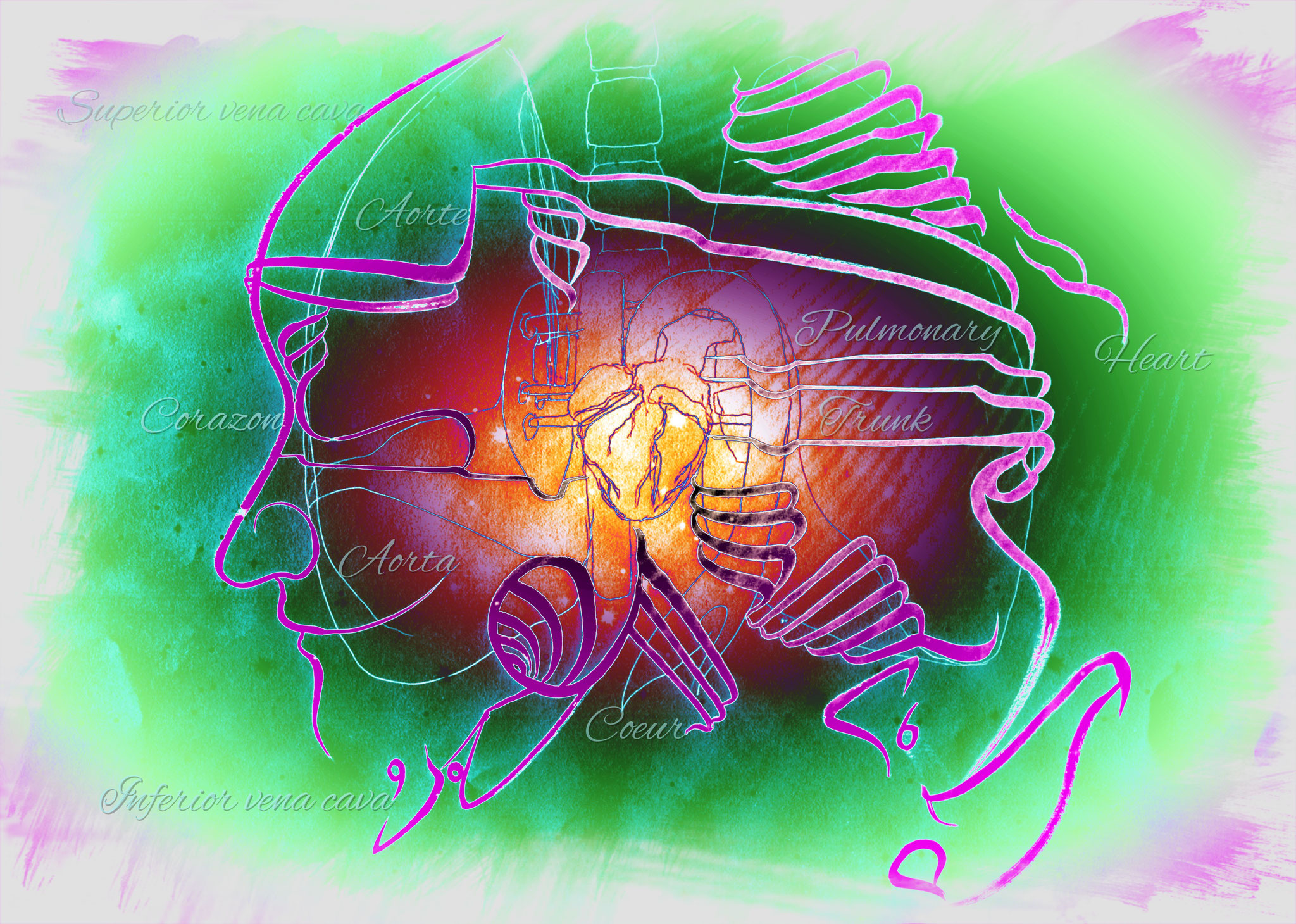
The King of Muscles
The heart is a pump formed of a spécial muscle, the heart muscle.
It is hollow and contains four cavities, two atria and two ventricles.
It contracts automatically with its muscle fibers by pressing blood.
Follow the current!
The blood passes from the right atrium to the right ventricle through the tricuspid valve.
Then he is sent to the lungs, where he picks up oxygen.
The red blood, rich in oxygen, goes from the lungs to the left atrium and then to the left ventricle.
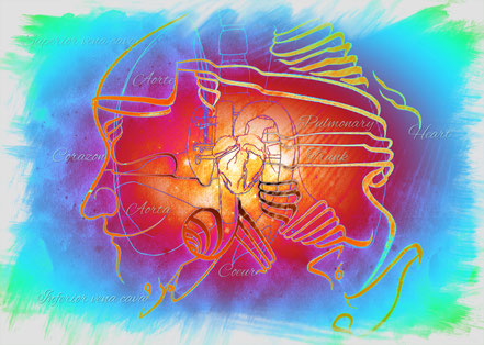
It is then sent in the aortic artery, to go everywhere in the body.
It can go to the brain through the carotid arteries, or to the arms through the axillary arteries, or continue to the legs via the femoral artery.
Finally, he returns to the starting point:
The heart!


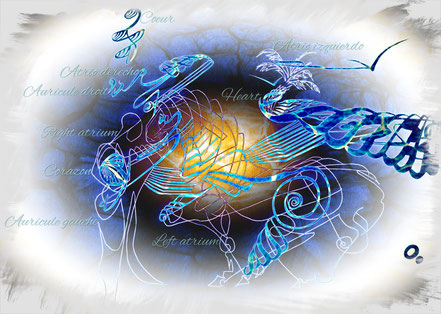

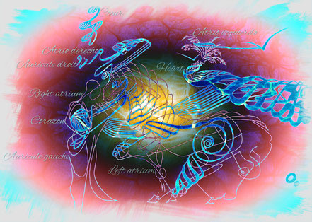
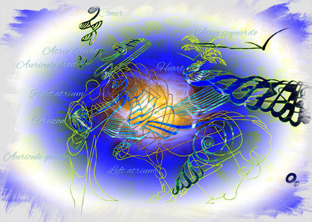


Right Auricule
The right atrium is one of the two small upper chambers of the heart, with a capacity of about 3 tablespoons.
This cavity receives all the venous blood(low in oxygen and saturated in carbon dioxide) brought by the vena cava,
As well as by the numerous small vesselswhich drain the walls of the cavity itself.
The right atrium is slightly more voluminous than the left atrium which is more powerful.
The walls of the right auricle, about 3millimeters thick, consist of two muscularlayers.
The superficial layer encompasses both atria, and the inner layer, composed of numerous small bundles, covers the atrial cavity at right angles to the superficiallayer.
During cardiac contraction, the blood is conducted into the pulmonary circulationvia the pulmonary valve.
When the heart relaxes, blood exits the rightatrium through the tricuspid valve to enter the right ventricle.
In the upper part of the right atrium, there is a small area of specialized cardiac tissue called the sino-atrial node:
It is the stimulator of the heart: it triggers and controls the contractions and the heart rythm.
The pulmonary trunk
It connects the right ventricle to the lungs!


The roads of the body
These roads are veins, arteries, veinlets and arterioles, to the finest paths: the capillaries, which is the central market, the place of all exchanges!
The arteries carry blood oxygenated from the heart to all other parts of the body.
They are lined with muscles to push blood along these "paths" to where it is requested!
The veins when they carry the blood loaded with impurities to the heart.
They are also lined with muscles allowing the dilation or contraction ensuring the management of the flow.
Because of their functions, the veins have "valves" that prevent reflux and forces the blood to flow in one direction!
Capillaries are the thinnest vessels that connect the arteries and veins.
They are so thin that the blood cells must almost run in single file to take these routes.
It is the place where nutrients, oxygen and various fluids pass through the tissue and where carbon dioxide and other waste
come to borrow to regain the circulation and leave the body.
"En route" to the head
From the aorta, the blood that goes towards the head borrows a large artery located at the level of the neck:
The carotid artery!


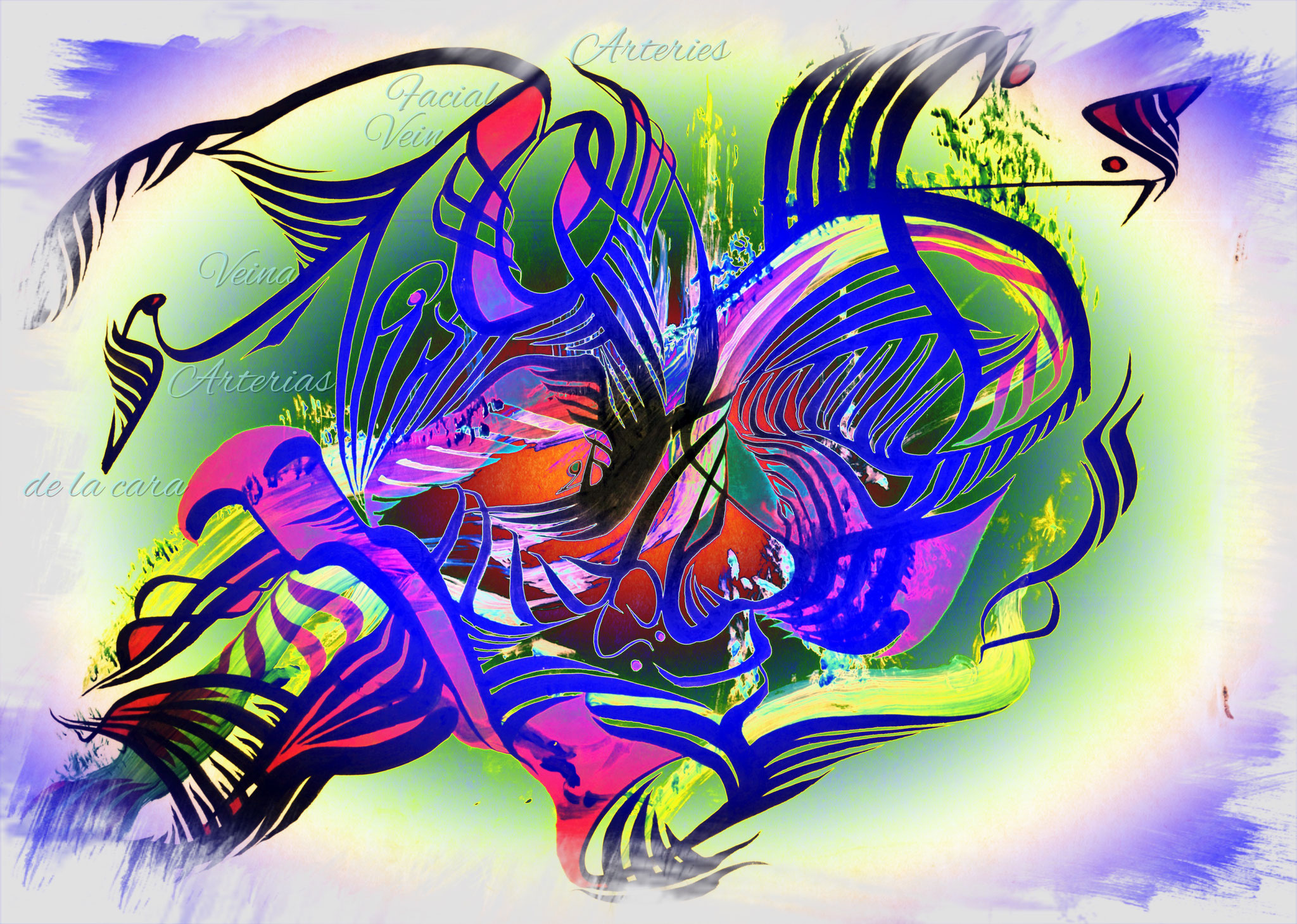
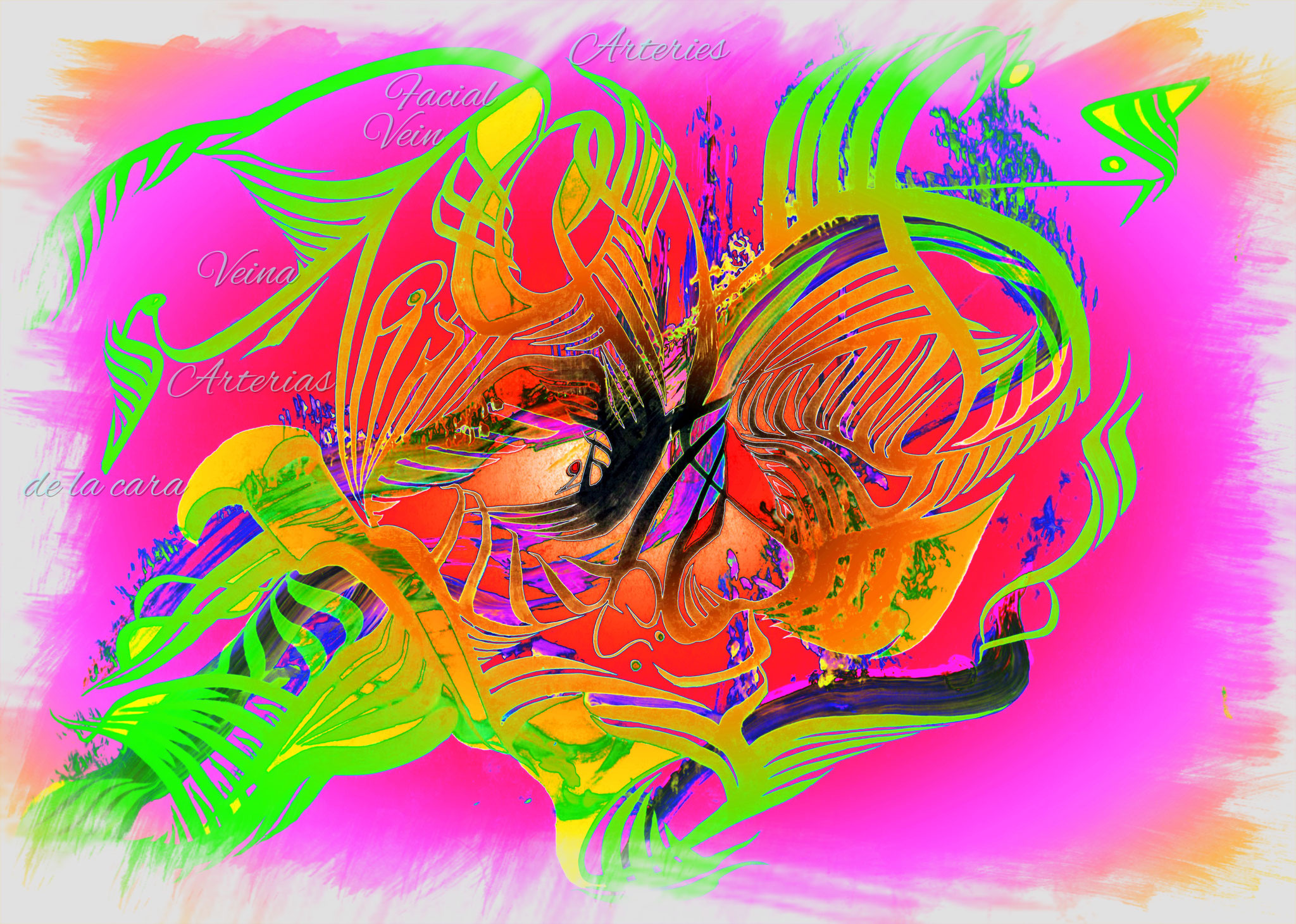
An irrigated smile
The arteries and veins of the face!
A simple title for a wave of oxygen!

All roads are open!
All along the body as well as in all our organs are veins and arteries, distributed like a network connecting the whole of the body to the heart.
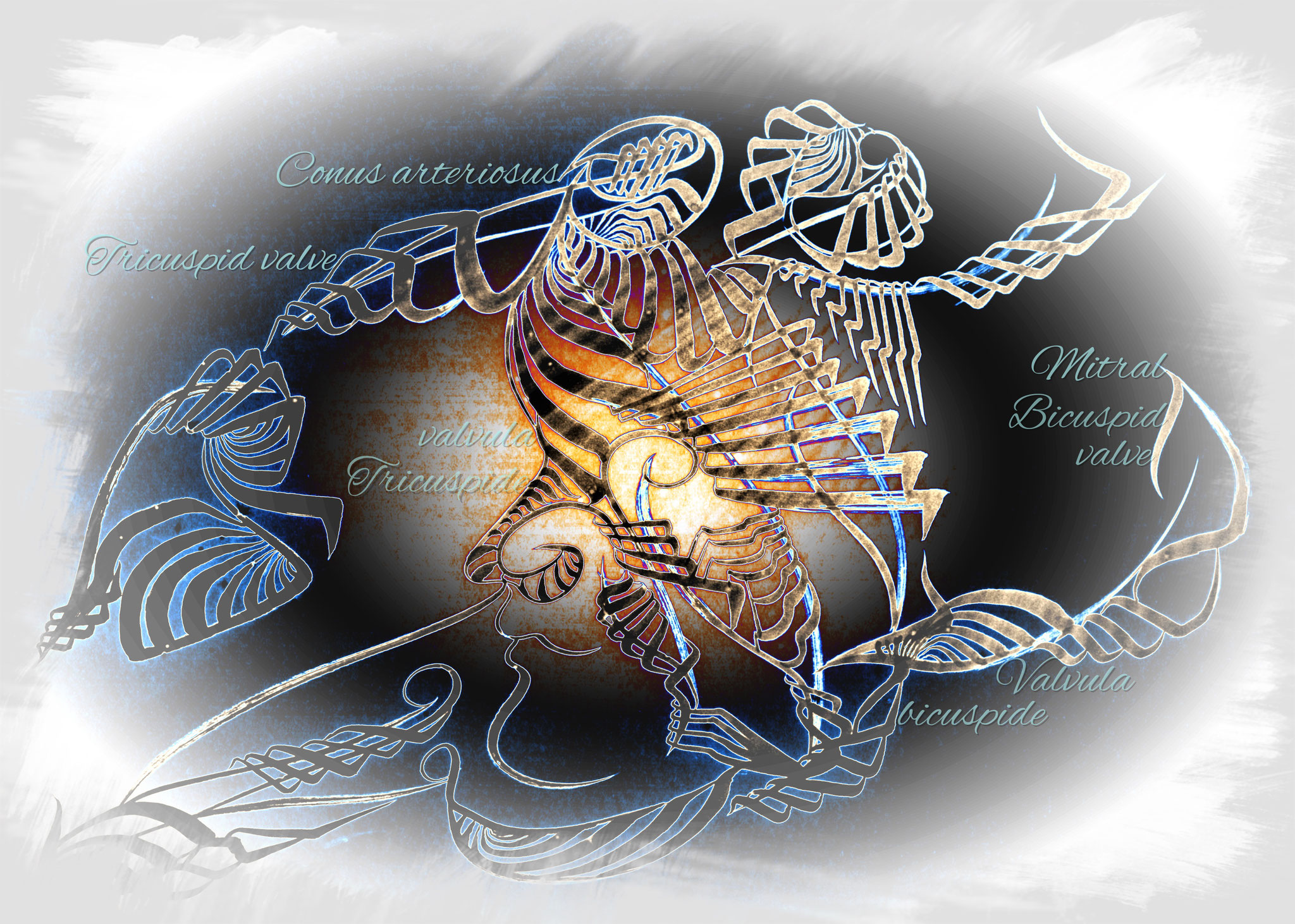
One heart, two valves
Heart valves are heart structures, separating the various cavities and preventing blood from flowing back in the wrong directions.
The tricuspid valve is a membranes fold consisting of three valves.
Located at the entrance of the passage separating the right ventricle from the right atrium of the heart.
The valvule located between the leftatrium and the left ventricle.
It consists of two mitral valves.
It is open during the filling of the left ventricle, leaving blood from the leftatrium passing freely.
During the contraction of the left ventricle, the mitral valves normally close, in a completely sealed manner.
The pulmonary valve is located at the entrance to the pulmonary artery and is constituted as the aorta of three sigmoid valves oriented towards the pulmonary artery.
It opens during contraction of the right ventricle, allowing the ejection of blood to the lungs.
Then it closes, avoiding the blood reflux.
The aortic valve located at the birth of the aorta is composed of three sigmoid valves in the form of cups open towards the aorta.
It opens because of blood pressure, during the contraction of the left ventricle.
This corresponds to the systole.
Then it closes during the release of the left ventricle, once the blood has been ejected into the aorta.